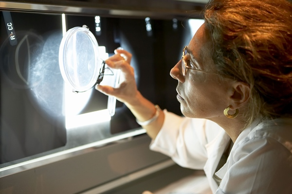
Get it early, get it right. When it comes to breast cancer, these two key points can make all the difference. The US-based non-profit National Public Radio (NPR) reported on a March 2015 study of pathologists in eight US states, where they were able to identify invasive breast cancer 96 per cent of the time, and normal tissue 87 per cent of the time.
However, the worrying bit was that ductal carcinoma in situ, the most common non-invasive type, and atypical hyperplasia, the accumulation of abnormal cells in the breast, were misdiagnosed 16 and a staggering 52 per cent of the time, respectively. “The first thing for women to remember is that making a diagnosis from tissue is part science and part art,” Dr Jean Simpson, President of Breast Pathology Consultants, told NPR.
No wonder many of the recent advances in breast cancer detection technologies have focused on making it more of a science and less of an art. This not only reduces mortality rates and patient trauma but also cuts down on false positives and unnecessary biopsies.
An inarguable fact, supported both by our regional experts on page 2 and by international research, is that breast cancer mortality has declined significantly over the past ten years — a lot of which has to do with early detection. Below, we take a look below at some new technologies that are aiding early diagnosis.
“The fact that you cannot argue with is that breast cancer mortality has declined by 24 per cent in the past 10 years, and a lot of that is due to early detection,” Dr Carol Lee, chairwoman of the Commission on Breast Imaging for the American College of Radiology, told the medical news and information source, WebMD.
3D mammography
Doctors can now peer deep inside the breast and generate high resolution 3D images of the tissue, layer by layer. A June 2014 report by the Journal of the American Medical Association reported a 41 per cent increase in detection of invasive cancer and 29 per cent increase in the detection of all breast cancers when 3D mammography was used. Washington Radiology Associates, an early adopter of this technology, saw an increased detection rate of 38 per cent.
3D-4D ultrasounds
Instead of radiation, high frequency sound is used to generate a detailed 3D map or a 4D live view of the breast tissue. The GE Logiq 9 with XDclear, for example, uses an array of advanced transducers that result in “extraordinary image quality”.
Elsewhere, the SoftVue from Delphinus Medical Technologies suspends the breast in a warm water bath while a transducer ring comprised of more than 2,000 elements emits ultrasound signals in a sequenced 360-degree circular array around it. The process takes just a couple of minutes. Unlike conventional mammograms, it is operator-independent and does not require painful breast compression.
Apparently, 3D ultrasounds are also better at differentiating between a cyst and a tumour.
Breast MRI
Instead of ultrasound or X-rays, MRI scans rely on powerful magnets and radio waves. A computer tracks resulting wave patterns to form a detailed image of the inside of a breast. A February 2015 study by the Medical University of Vienna’s radiology and nuclear medicine department reports around 90 per cent of all breast cancers can be identified using MRI — a sharp jump from the quoted 37.5 per cent for conventional mammography and ultrasound tests.
Meanwhile, in a September report, Radiology Today magazine reported on an experimental approach, using a non contrast-enhanced protocol, which cuts down the time to around seven minutes from the usual half-an-hour.
The future
Cancer.org, run by the American Cancer Society, lists emerging tech that may become the next big thing. Or may not. Tests are in the earliest stages of research and it will be a while before we know “if these imaging tests are as good as or better than those we use today”.
But among the promising ones is optical imaging, where light is focused on the breast — some of it passes through, while some of it bounces back, forming patterns that can help detect signs of cancer. This idea does not depend on breast compression or radiation. Researchers are also experimenting with merging optical imaging tech into existing tests like MRI or 3D mammography.
Another technology that is getting attention is Molecular Breast Imaging, which is a newer nuclear medicine imaging technique for the breast, and is suited for follow-up on breast problems. Also in contention is Positron Emission Mammography, which uses sugar attached to a radioactive particle to detect cancer cells. Apparently, this tech may be better at detecting small clusters of cancer cells within the breast


